
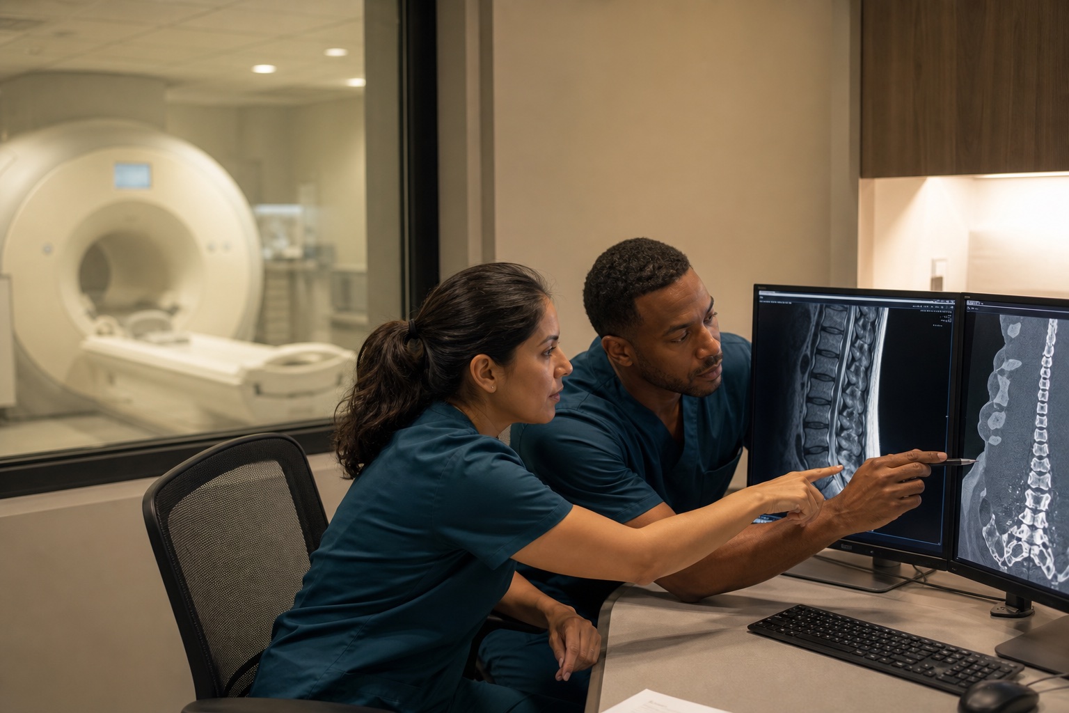
Radiology reports use terms like bulge, protrusion, extrusion, edema and degenerative change. Some findings suggest recent injury, while many are common with age. The radiologist describes the image; the treating physician connects it to the patient's history and exam. Neither should overstate what one image shows.
Who writes the MRI report and who interprets it?
A radiologist reads the images and writes the report. The radiologist usually has the order and a short clinical history but hasn't examined the patient. Their job is to describe what's visible as accurately as possible.
The treating physician, such as a pain management, spine or orthopedic specialist, examines the patient and decides how the findings relate to symptoms. Good care requires both. A report that says "disc protrusion at C5-6" means one thing in a patient with matching arm pain and weakness, and something else in a patient with no symptoms on that side.
What do the disc terms mean?
Radiologists use a standard vocabulary for disc findings, based on consensus nomenclature published by spine and radiology societies:
- Bulge: the disc extends evenly beyond the edges of the vertebrae over a wide area. Bulges are very common and often not a source of pain on their own.
- Protrusion: a focal area of disc material extends past the edge, with a base wider than its outward extent.
- Extrusion: disc material extends further, with the outward part wider than its base, or it has moved up or down.
- Sequestration: a fragment has separated from the disc.
- Annular fissure (sometimes called an annular tear): a separation in the outer fibers of the disc. Radiologists generally avoid implying a traumatic cause with this term alone.
Herniation is an umbrella term that covers protrusion, extrusion and sequestration.
Which findings can suggest a recent injury?
MRI can't date most findings precisely. Some features are more associated with recent injury:
- Bone marrow edema, seen as bright signal on fluid-sensitive sequences, which can indicate a bone bruise or acute fracture
- Ligament edema or disruption
- Soft tissue swelling or fluid
- An acute fracture line, sometimes with surrounding edema
- Disc height loss and desiccation (the disc appears darker due to less water)
- Osteophytes, or bone spurs
- Endplate changes that follow known chronic patterns
- Facet joint arthritis
- Ligament thickening
Some features are more associated with a long-standing process:
Many reports describe a mix. The presence of degeneration doesn't mean a patient wasn't injured, and the presence of a herniation doesn't mean it was caused by the incident. Those are clinical questions.
Why do so many people without pain have abnormal MRIs?
Published studies of people with no back pain have found disc bulges, degeneration and even protrusions in a large share of adults, increasing with age. That's why imaging alone doesn't establish a pain source or a cause. It's also why the history, exam, timing of symptoms and response to diagnostic procedures carry weight in the treating physician's assessment.
How does the treating physician connect imaging to symptoms?
Physicians look for concordance:
- Does the level and side of the finding match the patient's symptoms and exam?
- Do neurological signs, such as reflex changes, weakness or numbness in a specific pattern, fit the nerve root affected?
- Did symptoms begin or change after the incident, based on the history and prior records?
- How did the patient respond to targeted treatment, such as a selective nerve root block?
The physician's note should state that reasoning plainly, including when a finding is probably incidental.
Can comparing old and new MRIs help?
Yes, when prior imaging exists. A comparison can show whether a finding is new, unchanged or progressed. If a patient had earlier spine imaging, obtaining it with authorization is useful for the treating physician.
What should a reader avoid assuming from an MRI report?
- That "mild" findings can't cause symptoms, or that "severe" findings must
- That the word "acute" or "chronic" in the report settles causation
- That a normal MRI means there's no injury, since muscle, ligament and facet joint injuries often look normal
- That one MRI defines the patient's future
How does SpineLUX handle imaging?
Imaging is ordered when clinically indicated and coordinated through the network so results reach the ordering clinician. Reports and images are released with a valid HIPAA authorization. SpineLUX doesn't pay for referrals or promise any imaging finding or opinion.
What does an X-ray or CT add?
X-rays show bone alignment, fractures, disc space height and arthritis. Flexion and extension views can show abnormal movement between vertebrae. CT shows bone in fine detail and is the standard for suspected acute fractures. Neither shows discs, nerves or ligaments as well as MRI. Each study answers a different question, and the physician should note why it was ordered.
How should the treating physician's note discuss imaging?
A good note quotes or summarizes the main findings, states whether they match the exam, and explains which findings the physician considers clinically relevant and which are likely incidental. It should mention prior imaging if available. Short, specific reasoning reads better than a long list of every finding.
How should a reader handle conflicting reads?
Two radiologists sometimes describe the same study differently, one calling a finding a bulge and another a protrusion. That's common. The treating physician's note should explain which findings drive clinical decisions and why, rather than relying on a single word.
This article describes medical care and record practices only. It isn't legal advice and doesn't promise any clinical finding, opinion or case result.
Common questions
What else do patients ask?
Is a disc bulge an injury?+
A bulge is a description, not a diagnosis. Whether it relates to symptoms is a clinical judgment.
Can MRI show how old a herniation is?+
Not precisely. Some features suggest recency or chronicity, but timing usually depends on history and prior imaging.
What is bone marrow edema?+
Fluid signal in bone that can indicate a bruise, fracture or other process. Its meaning depends on location and context.
Why would a physician order a second MRI?+
To evaluate new or changing symptoms, or to plan a procedure. It's ordered for a clinical reason.
Does upright or open MRI change the findings?+
Different machines and positions can show findings differently. Physicians interpret each study in context.