
There is no single best scan
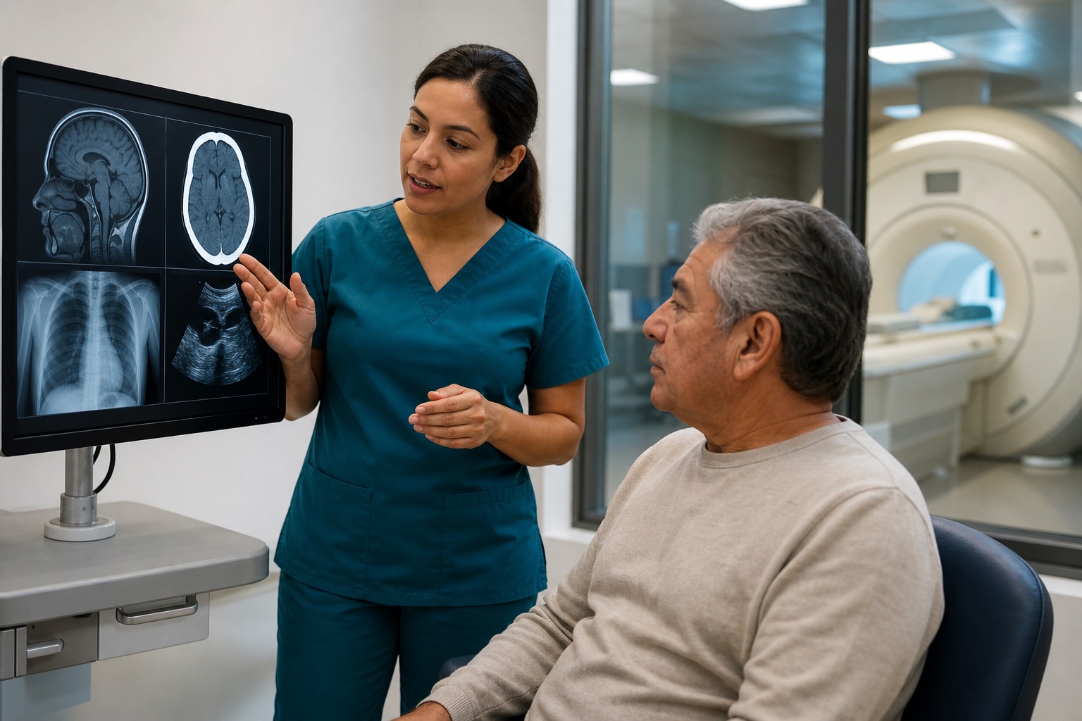
X-ray, CT, MRI, and ultrasound use different technologies and reveal different tissues. The right choice depends on what the clinician is trying to find, how urgent the situation is, and whether radiation, contrast, metal implants, pregnancy, or other safety considerations apply.
Imaging is most useful when it follows a focused history and examination. A scan should help answer a question—not replace clinical judgment.
X-ray: bones and alignment
X-rays are commonly used for suspected fractures, dislocations, joint alignment, and certain degenerative changes. They are quick and widely available, which makes them useful in urgent settings.
Their limitation is soft-tissue detail. Muscles, tendons, ligaments, discs, nerves, and cartilage are not shown as clearly as bone. A normal X-ray can be reassuring without answering every question about ongoing pain or instability.
CT: detailed cross-sectional trauma imaging
Computed tomography uses X-rays and computer processing to create cross-sectional images. CT is valuable for complex fractures, certain head and spine emergencies, chest or abdominal trauma, and situations where speed matters.
CT uses ionizing radiation, generally more than a standard X-ray. That benefit can outweigh exposure when serious injury is suspected. Contrast may be used, so share allergy, kidney, pregnancy, and medication information with the care team.
MRI: soft tissue, brain, spine, and nerves
MRI uses a strong magnetic field and radiofrequency energy rather than ionizing radiation. It can show the brain, spinal cord, discs, nerves, ligaments, tendons, cartilage, bone marrow, and many joint structures in detail.
MRI takes longer than CT and requires screening for implanted devices, metal fragments, claustrophobia, and other safety issues. It is not automatically the first test after every accident, but it can be valuable when soft-tissue or neurological injury is suspected.
Ultrasound and DTI
Ultrasound uses sound waves and can evaluate certain tendons, muscles, ligaments, fluid collections, blood flow, and superficial structures in real time. It does not use ionizing radiation, but it is not ideal for every brain, spine, lung, or deep-joint question.
Diffusion tensor imaging, or DTI, is an MRI-based technique that measures water-diffusion patterns in white matter. It may be considered in selected neurological contexts, but it should not be treated as stand-alone proof of traumatic brain injury. SpineLUX considers DTI/MRI alongside neurological history, examination, and other findings.
Questions to ask before imaging
Good imaging decisions are collaborative. Ask what the study should evaluate, whether the result could change treatment, what safety screening is needed, and who will explain the report.
- What specific clinical question should this test answer?
- Does the test use radiation or contrast?
- Could implants, pregnancy, kidney disease, or claustrophobia affect the plan?
- Who will connect the imaging result to treatment?
Common questions
Frequently asked questions
Is MRI better than CT after a car accident?+
Neither is universally better. CT is fast and strong for many acute trauma and bone questions; MRI provides greater soft-tissue and neurological detail.
Does ultrasound use radiation?+
No. Ultrasound uses sound waves. MRI also does not use ionizing radiation, while X-ray and CT do.