
Begin with the neurological question
A diagnostic test is most useful when it is ordered to answer a focused clinical question. After a head injury, that question may involve episodes of altered awareness, seizure-like activity, unexplained confusion, persistent cognitive symptoms, sleep-related events, or another neurological concern. The patient’s history and neurological examination come first.
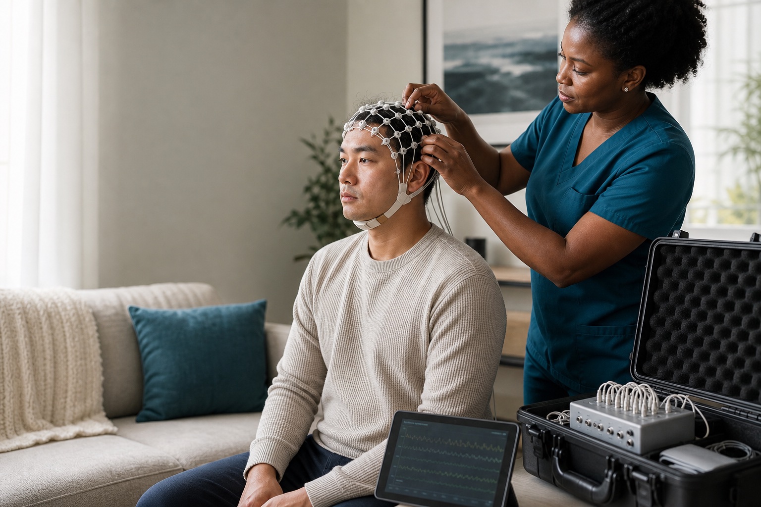
SpineLUX offers telehealth or in-clinic neurological consultation and can coordinate mobile EEG, mobile qEEG, and mobile brain mapping across Texas when clinically appropriate. Mobile availability means the equipment and trained personnel can travel to an eligible setting; it does not mean every patient automatically needs all three services.
What an EEG records
An electroencephalogram, or EEG, records electrical activity detected at the scalp through small electrodes. A routine EEG can help a clinician evaluate patterns associated with seizures and other disturbances of brain function. Longer or specialized recordings may be considered when brief testing is unlikely to capture the event of concern.
EEG does not take a picture of the brain. It measures electrical signals over time. A normal routine EEG does not rule out every seizure disorder, concussion, or neurological condition. Likewise, an abnormal pattern must be interpreted by a qualified clinician in the context of symptoms, medications, medical history, and technical quality.
How qEEG and brain mapping relate to EEG
Quantitative EEG, commonly called qEEG, applies mathematical and statistical analysis to recorded EEG data. Brain mapping is a visual way of displaying selected qEEG measurements across scalp regions. These tools may help a qualified clinician review patterns, compare regions, or support specific forms of assessment.
The American Clinical Neurophysiology Society has cautioned that qEEG is not a stand-alone diagnostic test for mild traumatic brain injury. Results can be influenced by artifact, drowsiness, medication, age, recording conditions, and the methods used for analysis. A map can look precise while still requiring careful clinical interpretation.
Mobile service can reduce access barriers
Travel can be difficult for a person experiencing headaches, dizziness, pain, anxiety, cognitive fatigue, or transportation problems after an injury. Mobile neurodiagnostic service can bring appropriate equipment to an eligible clinic, office, or other approved setting and may help attorneys, families, and care teams coordinate evaluation across distant Texas markets.
Before scheduling, the team must confirm the clinical order, location, electrical and space requirements, patient mobility, recording duration, and whether the setting can support a technically adequate study. Some clinical situations still require a hospital, epilepsy monitoring unit, emergency department, or another facility with immediate medical support.
Information that supports a useful referral
A complete referral helps the neurologist understand why testing is being considered. It should state the clinical question rather than simply request a device or map. Describe the event, symptom timeline, witnessed episodes, head impact or loss of awareness, emergency findings, current medications, previous neurological conditions, and relevant imaging or prior EEG results.
Do not send protected health information through ordinary email. SpineLUX provides a secure attorney referral form and can guide referring offices to the correct clinical pathway. Patient authorization, record-release requirements, and the minimum necessary information should be respected.
- Date and mechanism of injury or onset of symptoms
- Description, duration, and frequency of concerning episodes
- Witness observations, emergency records, and relevant examination findings
- Current medications and prior neurological or seizure history
- Existing CT, MRI, DTI, EEG, or specialist records
- Language, mobility, transportation, and location needs
EEG and MRI answer different questions
EEG measures electrical activity. CT and MRI evaluate anatomy and structural findings. Diffusion tensor imaging, or DTI, is an MRI technique that analyzes the direction of water movement in white-matter pathways. These studies are not interchangeable, and none should be ordered simply because another test was normal.
The American College of Radiology publishes appropriateness criteria for head trauma imaging. The selected study depends on the timing, symptoms, examination, and suspected condition. A clinician may decide that structural imaging, electrical recording, both, or neither is appropriate for a particular patient.
What patients can expect
The scheduling team will confirm preparation instructions, medications, hair and scalp requirements, arrival time, and the expected length of the recording. Patients should follow the specific instructions they receive; they should not stop prescribed medication unless the ordering clinician directs them to do so.
During a standard recording, electrodes are placed on the scalp with conductive material. The study is generally noninvasive and records activity; routine electrodes do not deliver electricity into the brain. After technical processing, a qualified clinician interprets the record and places the findings within the broader neurological evaluation.
A responsible report states both findings and limits
A high-quality neurodiagnostic process includes proper acquisition, artifact review, qualified interpretation, and a report that answers the referral question without overstating certainty. Results should be shared with the treating team so they can be considered alongside symptoms, function, examination, and other studies.
No EEG, qEEG, brain map, MRI, or DTI result should be used alone to promise a traumatic-brain-injury diagnosis, determine legal causation, or predict a case outcome. For attorneys and case managers, the most reliable approach is to facilitate access, maintain authorized records, and allow the clinician to determine medical necessity and interpretation.
Common questions
Frequently asked questions
Are EEG, qEEG, and brain mapping available as mobile services?+
Yes. SpineLUX can coordinate mobile EEG, mobile qEEG, and mobile brain mapping across Texas when clinically appropriate and when the proposed setting can support a technically adequate study.
Can qEEG diagnose a concussion by itself?+
No. Professional guidance does not support qEEG as a stand-alone diagnostic test for mild traumatic brain injury. Interpretation must be integrated with the clinical history, examination, and other appropriate studies.
Is a neurology consultation available before testing?+
Yes. Neurological consultation may be available through telehealth or an in-clinic visit. The clinician can determine whether EEG, qEEG, brain mapping, imaging, or another pathway is appropriate.