
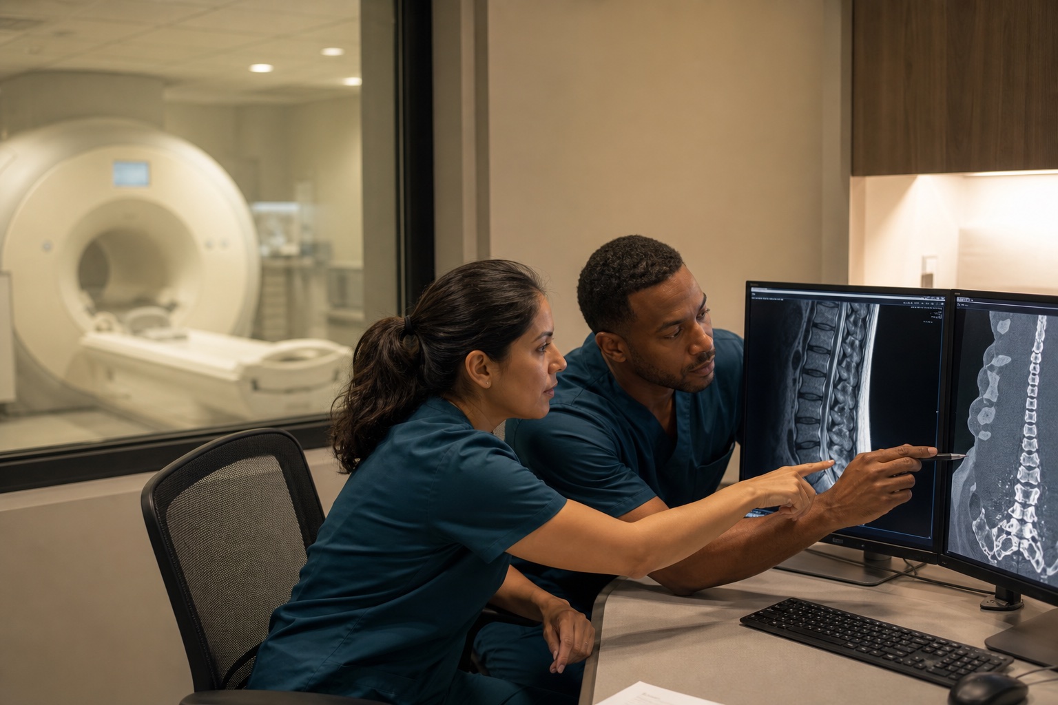
Imaging should answer a clinical question
The purpose of imaging is not simply to produce a report. It is to clarify anatomy, evaluate a suspected injury, guide treatment, or rule out a concern raised by the history and examination. The most efficient imaging pathway begins with a specific question from the treating clinician.
For example, an X-ray may evaluate fracture or alignment. CT can provide fast, detailed trauma and bone imaging. MRI can show discs, nerves, the spinal cord, brain, ligaments, tendons, cartilage, and other soft tissues. Ultrasound can evaluate selected structures dynamically without ionizing radiation.
The right test depends on the structure and urgency
No modality is best for every injury. CT may be preferred in an emergency because speed matters. MRI may be more useful for a persistent neurological or soft-tissue question. Ultrasound can be valuable for a superficial tendon, muscle, or fluid collection. DTI/MRI may be considered in selected neurological contexts.
Safety also matters. The team should consider radiation, contrast, pregnancy, kidney function, implanted devices, metal exposure, and claustrophobia. Prior imaging should be compared whenever possible.
A report is not the end of the process
Radiologists interpret images and describe findings, but treating clinicians connect those findings to the patient. Some abnormalities are old or incidental. Some meaningful injuries are subtle. The specialist must decide whether the imaging matches the symptom location, examination, and functional changes.
Timely report review matters in injury care. If a study identifies a fracture, significant disc problem, tendon tear, or another actionable finding, the referral and treatment plan should move without requiring the patient to coordinate every handoff alone.
Coordinated imaging reduces delay and duplication
When imaging and specialists operate in isolation, patients may repeat studies, carry discs between offices, or wait for one provider to learn that another already found the answer. Shared clinical questions and organized record flow can make the next visit more productive.
SpineLUX coordinates imaging with pain management, orthopedics, spine care, and neurology so the result can inform care efficiently. Imaging remains diagnosis-led and based on individual need.
Common questions
Frequently asked questions
Which imaging test is best after an accident?+
The best test depends on the suspected structure, urgency, examination findings, and safety considerations. No single modality is best for every injury.
Why should a specialist review the imaging report?+
The treating specialist connects imaging findings with symptoms, examination, prior studies, and the treatment decision.